

Parametry
Więcej o książce
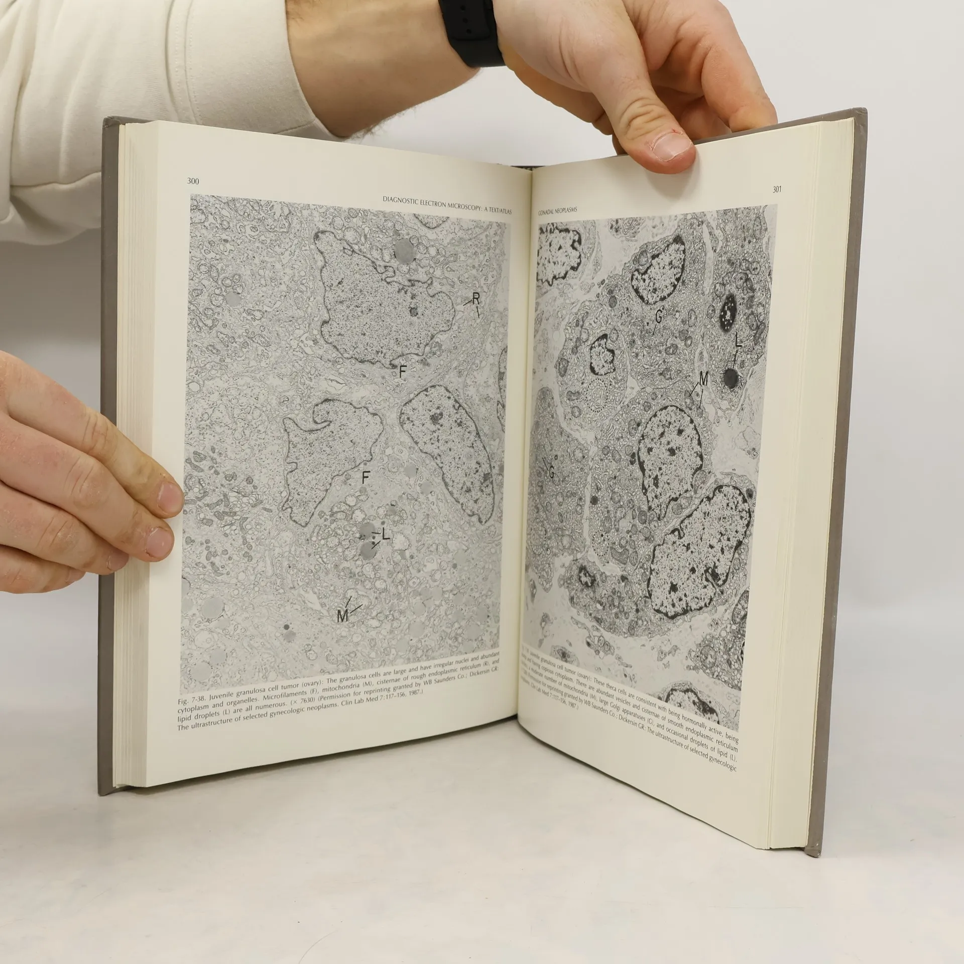
This text atlas, now in its second edition, presents in simplest form the basic diagnostic criteria used by the electron microscopist in studying neoplasms and other diseases encountered in the routine practice of pathology. Every field of electron microscopy is covered and low magnification plates are juxtaposed with higher magnifications to illustrate diagnostic features. The largest section of the book is devoted to neoplasms as this is the area in which most diagnostic problems occur. Renal glomerular disease is another important category in which ultrastructural study may be critical in diagnosis; infectious diseases, especially those of viral, protozoan, and unusual bacterial etiologies, are a third area in which electron microscopy may be used to establish or susbstantiate a diagnosis. All of these areas are comprehensively covered with concise, readable text and more than 800 first-quality images. This book is the preeminent reference for pathologists needing current information on the role of ultrastructure in diagnostic pathology.
Zakup książki
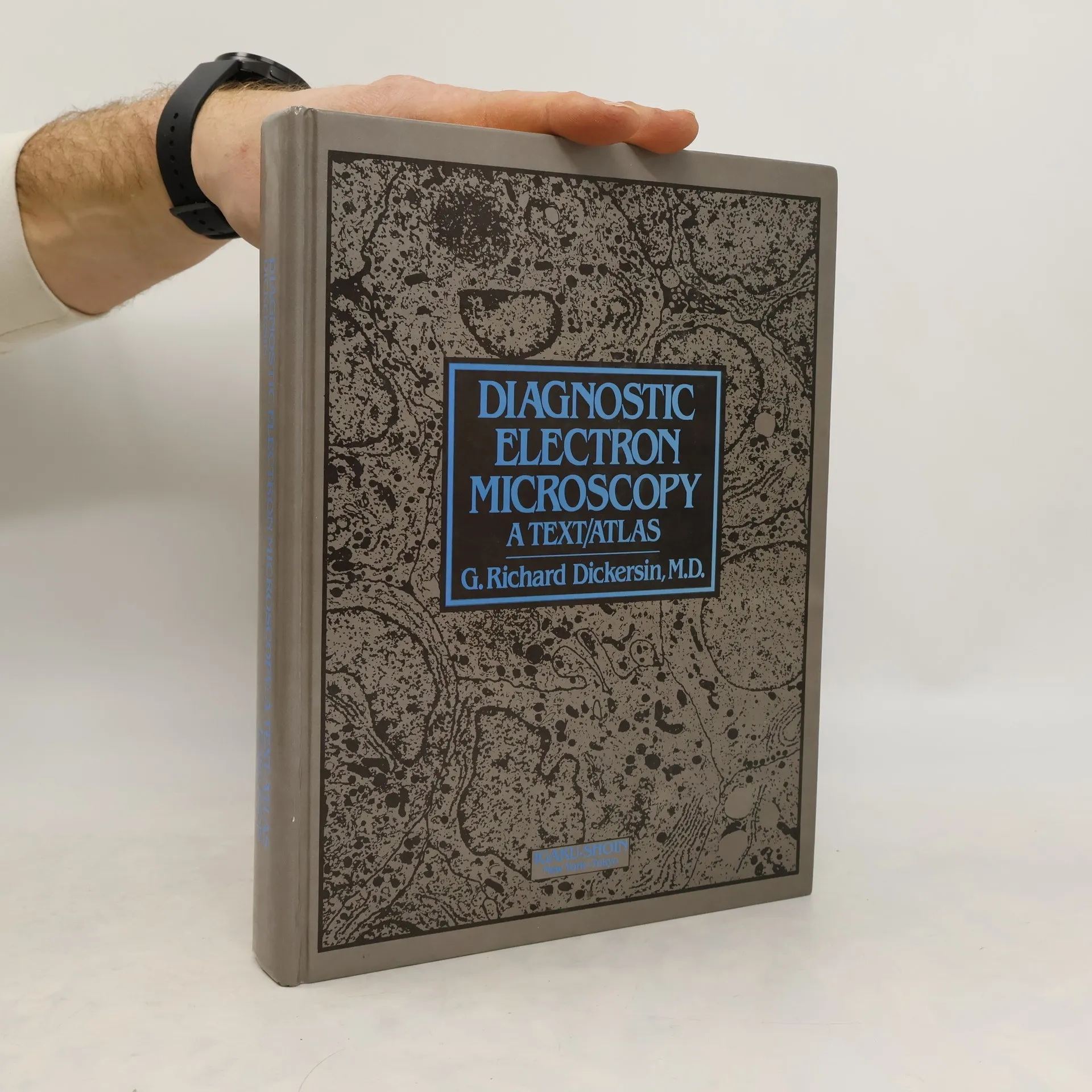
Diagnostic Electron Microscopy, G. Richard Dickersin
- Język

- Rok wydania
- 1988
- Oprawa
- (twarda)
Metody płatności
Nikt jeszcze nie ocenił.
- Tytuł
- Diagnostic Electron Microscopy
- Język
- angielski
- Autorzy
- G. Richard Dickersin
- Rok wydania
- 1988
- Oprawa
- twarda
- ISBN10
- 0896401375
- ISBN13
- 9780896401372
- Seria
- Kategorie
- Opis
- This text atlas, now in its second edition, presents in simplest form the basic diagnostic criteria used by the electron microscopist in studying neoplasms and other diseases encountered in the routine practice of pathology. Every field of electron microscopy is covered and low magnification plates are juxtaposed with higher magnifications to illustrate diagnostic features. The largest section of the book is devoted to neoplasms as this is the area in which most diagnostic problems occur. Renal glomerular disease is another important category in which ultrastructural study may be critical in diagnosis; infectious diseases, especially those of viral, protozoan, and unusual bacterial etiologies, are a third area in which electron microscopy may be used to establish or susbstantiate a diagnosis. All of these areas are comprehensively covered with concise, readable text and more than 800 first-quality images. This book is the preeminent reference for pathologists needing current information on the role of ultrastructure in diagnostic pathology.