

Parametry
Więcej o książce
Ultrasonography is an important adjuvant for the clinical assessment of a variety of ocular and orbital diseases. With proper use, one can gather a vast amount of information not possible with physical exam alone. Ultrasound is most useful when intraocular are difficult or impossible to examine. Situations that prevent normal examination include lid problems (severe edema, partial or total tarsorrhaphy), keratoprosthesis, corneal opacities, hyphema, hypopyon, miosis, pupillary membranes, dense cataracts, or vitreous opacities (hemorrhage, inflammatory debris). Diagnostic B-scan ultrasound accurately images intraocular structures and give valuable information on the status of the lens, vitreous, retina, choroid, and sclera. Ultrasound is also used for diagnostic purposes when pathology is clinically visible, such as differentiating iris or ciliary body lesions; ruling out ciliary body detachments; differentiating intraocular tumors, serous versus hemorrhagic choroidal detachments, rhegmatogenous versus exudative retinal detachments, and disc drusen versus papilledema; or determining functioning versus nonfunctioning glaucoma tube shunts.
Zakup książki
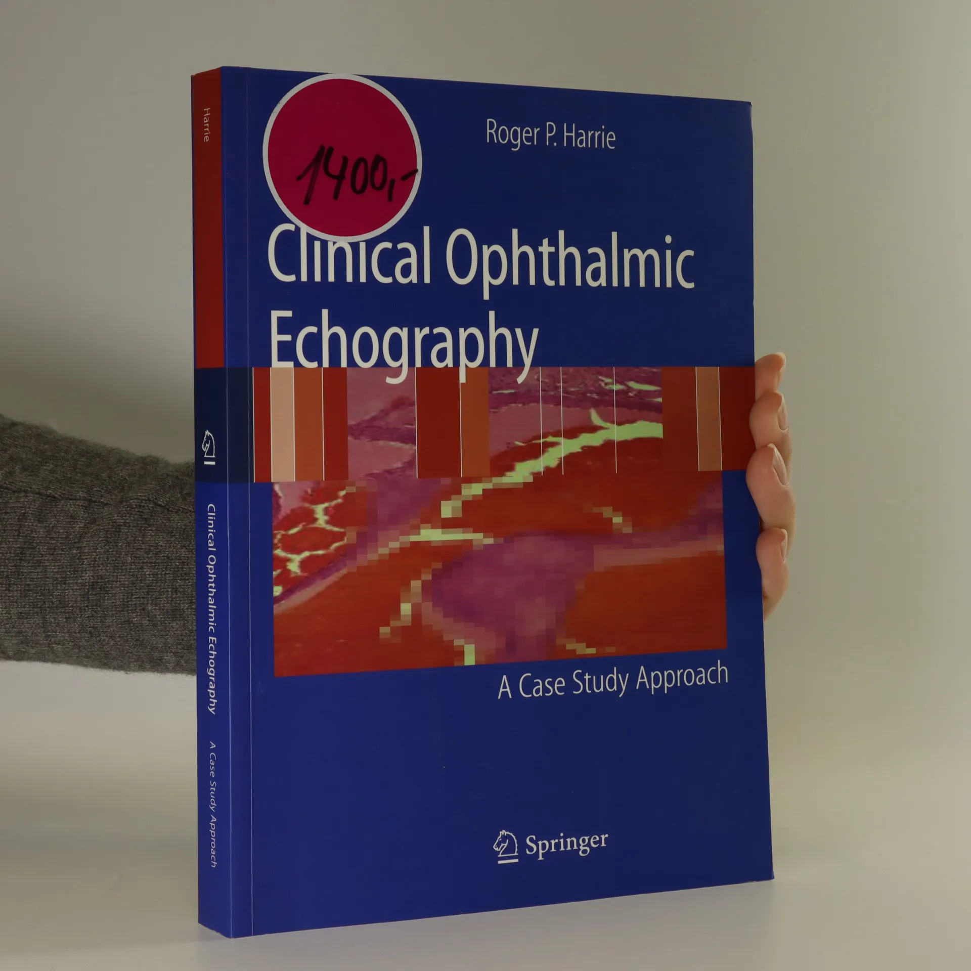
Clinical Ophthalmic Echography A Case Study Approach, Roger P. Harrie
- Język

- Rok wydania
Metody płatności
Nikt jeszcze nie ocenił.
- Tytuł
- Clinical Ophthalmic Echography A Case Study Approach
- Język
- angielski
- Autorzy
- Roger P. Harrie
- Wydawca
- Springer
- ISBN10
- 0387752447
- ISBN13
- 9780387752440
- Seria
- Kategorie
- Opis
- Ultrasonography is an important adjuvant for the clinical assessment of a variety of ocular and orbital diseases. With proper use, one can gather a vast amount of information not possible with physical exam alone. Ultrasound is most useful when intraocular are difficult or impossible to examine. Situations that prevent normal examination include lid problems (severe edema, partial or total tarsorrhaphy), keratoprosthesis, corneal opacities, hyphema, hypopyon, miosis, pupillary membranes, dense cataracts, or vitreous opacities (hemorrhage, inflammatory debris). Diagnostic B-scan ultrasound accurately images intraocular structures and give valuable information on the status of the lens, vitreous, retina, choroid, and sclera. Ultrasound is also used for diagnostic purposes when pathology is clinically visible, such as differentiating iris or ciliary body lesions; ruling out ciliary body detachments; differentiating intraocular tumors, serous versus hemorrhagic choroidal detachments, rhegmatogenous versus exudative retinal detachments, and disc drusen versus papilledema; or determining functioning versus nonfunctioning glaucoma tube shunts.