

Więcej o książce
The demand for an atlas of CSF cytology has made a second edition necessary very soon. The number of those who have learned to appreciate the value of this simple diagnostic procedure has apparently increased. Optimal methods of cell preparation, lim ited technical requirements and immediate availabil ity of the results have all contributed to this develop ment. In the current edition, primary emphasis has again been placed on selection of informative illustrations and on quality of reproduction. Some figures have been replaced, and some added. The text has been expanded and improved where it seemed appro priate. Malignant lymphomas are treated as a sepa rate topic, rather than as part of the chapter on he moblastoses. I should like to thank Prof. Dr. HANSJORG RIEHM of the pediatric department at Kli nikum Charlottenburg, Freie UniversiHit Berlin (Director: Prof. Dr. HANS HELGE), for his suggestion in this regard, as well as for the material for figures on plate 47 (a-d). My technical assistent, Mrs. CHRISTINE LIGNER, again deserves my special thanks. Her suggestions and her careful cytologic work have been significant contributions to the success of this atlas.
Zakup książki
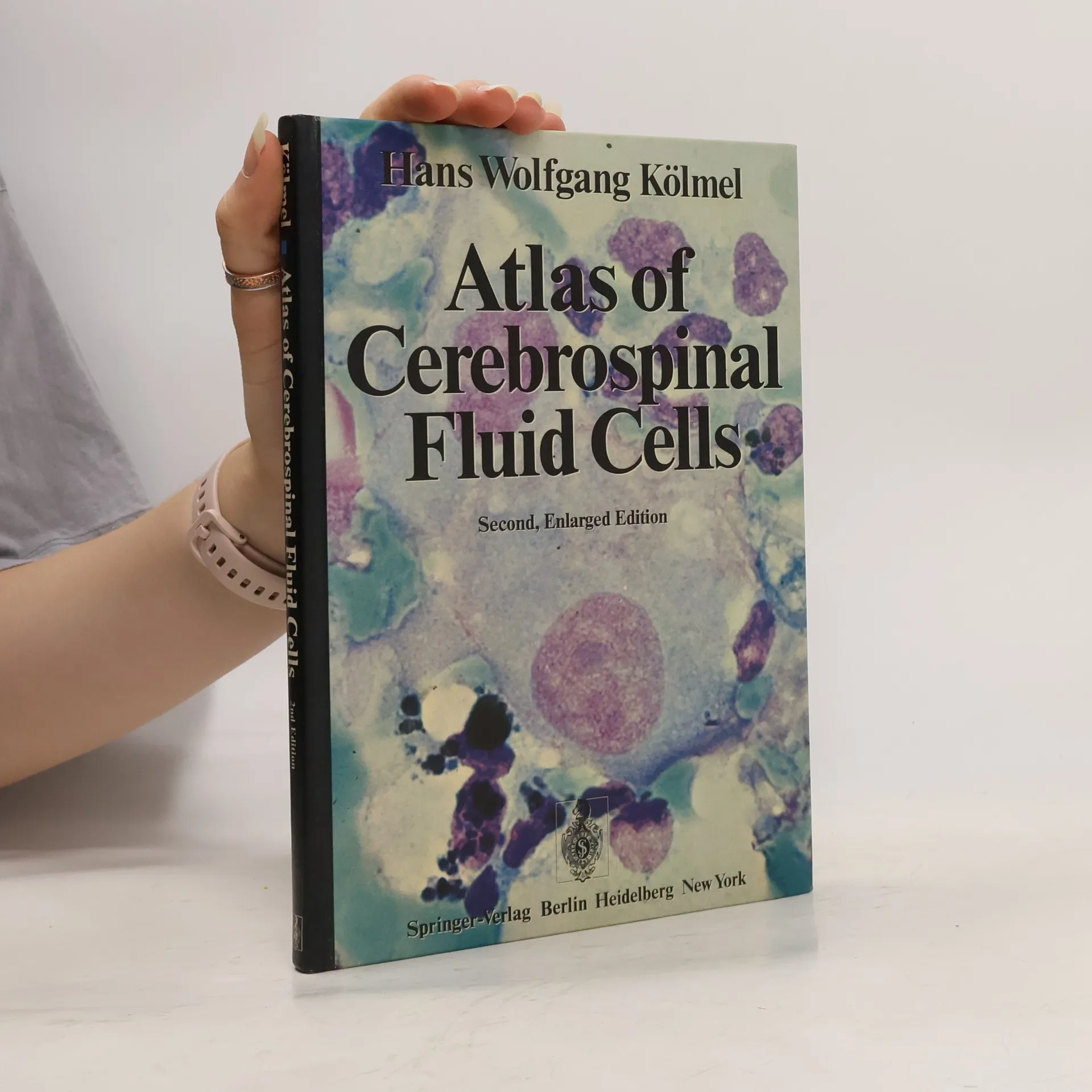
Atlas of Cerebrospinal Fluid Cells - Second, Enlarged Edition, Hans Wolfgang Kölmel
- Język

- Rok wydania
- 1977
- Oprawa
- (twarda)
Metody płatności
Nikt jeszcze nie ocenił.
- Tytuł
- Atlas of Cerebrospinal Fluid Cells - Second, Enlarged Edition
- Język
- angielski
- Autorzy
- Hans Wolfgang Kölmel
- Rok wydania
- 1977
- Oprawa
- twarda
- Liczba stron
- 144
- ISBN10
- 3540081860
- ISBN13
- 9783540081869
- Seria
- Opis
- The demand for an atlas of CSF cytology has made a second edition necessary very soon. The number of those who have learned to appreciate the value of this simple diagnostic procedure has apparently increased. Optimal methods of cell preparation, lim ited technical requirements and immediate availabil ity of the results have all contributed to this develop ment. In the current edition, primary emphasis has again been placed on selection of informative illustrations and on quality of reproduction. Some figures have been replaced, and some added. The text has been expanded and improved where it seemed appro priate. Malignant lymphomas are treated as a sepa rate topic, rather than as part of the chapter on he moblastoses. I should like to thank Prof. Dr. HANSJORG RIEHM of the pediatric department at Kli nikum Charlottenburg, Freie UniversiHit Berlin (Director: Prof. Dr. HANS HELGE), for his suggestion in this regard, as well as for the material for figures on plate 47 (a-d). My technical assistent, Mrs. CHRISTINE LIGNER, again deserves my special thanks. Her suggestions and her careful cytologic work have been significant contributions to the success of this atlas.