



Parametry
- 106 stron
- 4 godziny czytania
Więcej o książce
This book is written by an author whose knowledge of the subject is based on vast surgical experience. A reliable assessment of the success or failure of a micro vascular anastomosis or an aneurysm operation has until now been possible only postoperatively by angiography and transcutaneous Doppler sonography. Of particular impor tance are the stenoses and occlusions which can occur dur ing the operation, but which are frequently not detected un til after surgery. This induced the neurosurgeons in Freiburg, as well as those in several other hospitals, to carry out a rou tine control with postoperative transcutaneous Doppler fol lowing extracranial-intracranial bypass operations and an giography while still under anesthesia following aneurysm operations. The stenoses and occlusions detected were often not apparent intraoperatively. The reasons for this are that from the outside we see only the surface - and not the lu men - of a v ssel, and that stenoses caused by thrombosis, indentation of the vessel wall during anastomosis or by plaque on the vessel wall are not externally obvious. Even arterial pulsation is no guarantee of patency. The necessity to be able to detect stenoses during the op eration is obvious. The microvascular Doppler sonography adopted by Dr. Gilsbach provides an expedient and safe method of recognizing such lumen narrowings.
Zakup książki
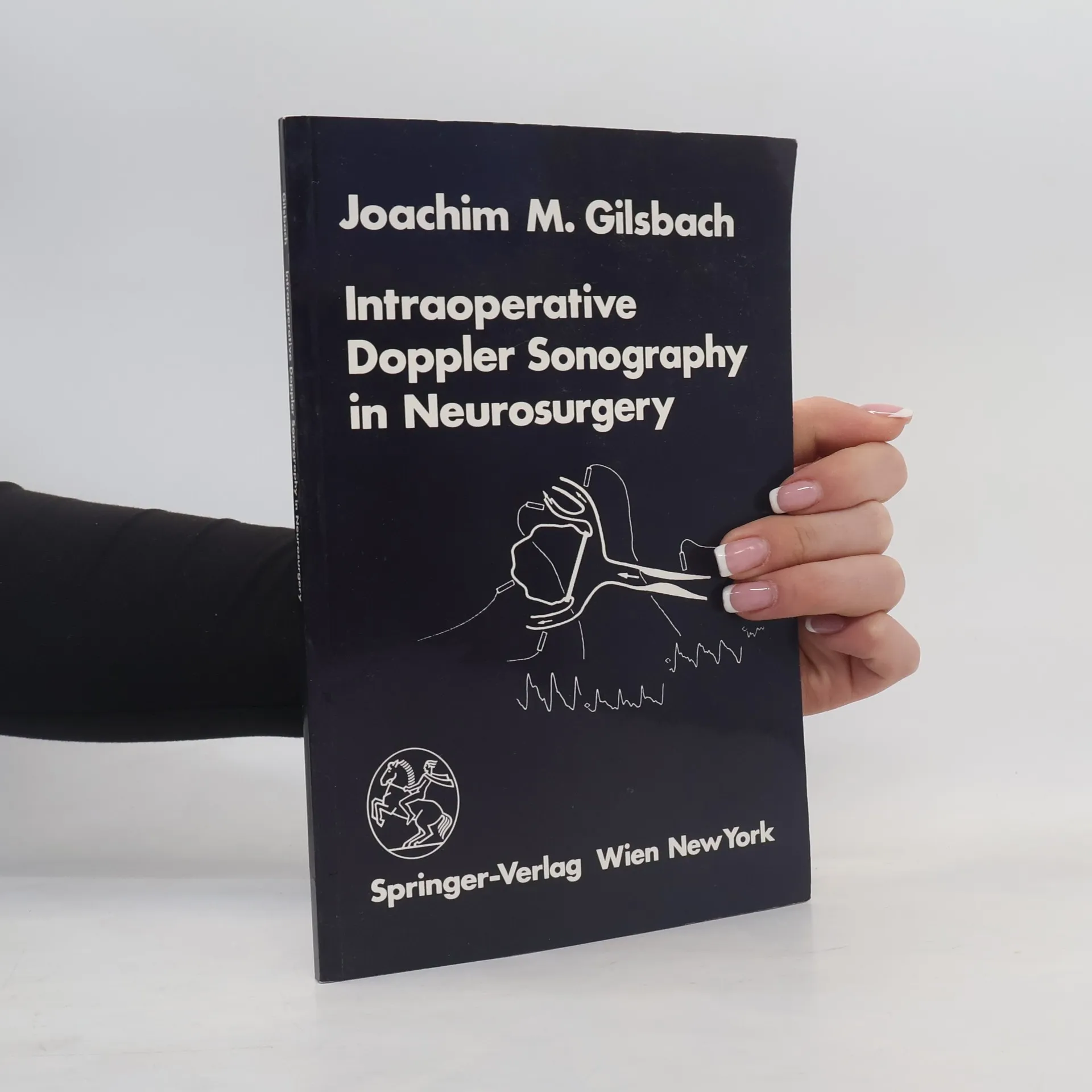
Intraoperative Doppler Sonography in Neurosurgery, Joachim M. Gilsbach
- Język

- Rok wydania
- 1983
- Oprawa
- (miękka)
Metody płatności
Brakuje nam tutaj Twojej recenzji.
- Tytuł
- Intraoperative Doppler Sonography in Neurosurgery
- Język
- angielski
- Autorzy
- Joachim M. Gilsbach
- Wydawca
- Springer
- Rok wydania
- 1983
- Oprawa
- miękka
- Liczba stron
- 106
- ISBN10
- 3211817689
- ISBN13
- 9783211817681
- Seria
- Kategorie
- Ocena
- 5 z 5
- Opis
- This book is written by an author whose knowledge of the subject is based on vast surgical experience. A reliable assessment of the success or failure of a micro vascular anastomosis or an aneurysm operation has until now been possible only postoperatively by angiography and transcutaneous Doppler sonography. Of particular impor tance are the stenoses and occlusions which can occur dur ing the operation, but which are frequently not detected un til after surgery. This induced the neurosurgeons in Freiburg, as well as those in several other hospitals, to carry out a rou tine control with postoperative transcutaneous Doppler fol lowing extracranial-intracranial bypass operations and an giography while still under anesthesia following aneurysm operations. The stenoses and occlusions detected were often not apparent intraoperatively. The reasons for this are that from the outside we see only the surface - and not the lu men - of a v ssel, and that stenoses caused by thrombosis, indentation of the vessel wall during anastomosis or by plaque on the vessel wall are not externally obvious. Even arterial pulsation is no guarantee of patency. The necessity to be able to detect stenoses during the op eration is obvious. The microvascular Doppler sonography adopted by Dr. Gilsbach provides an expedient and safe method of recognizing such lumen narrowings.