


Więcej o książce
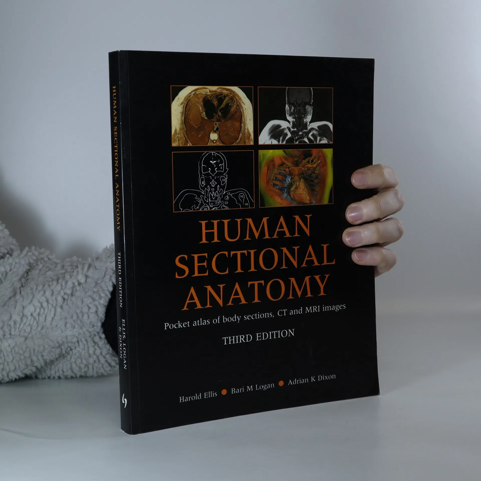
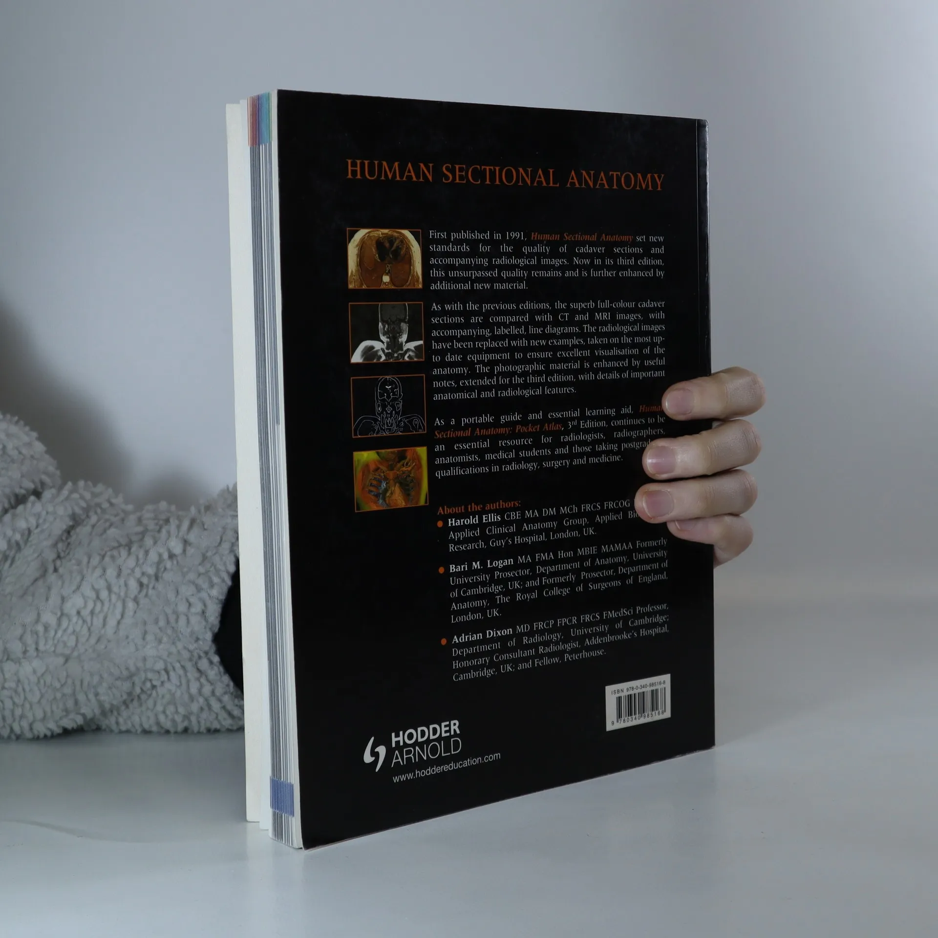
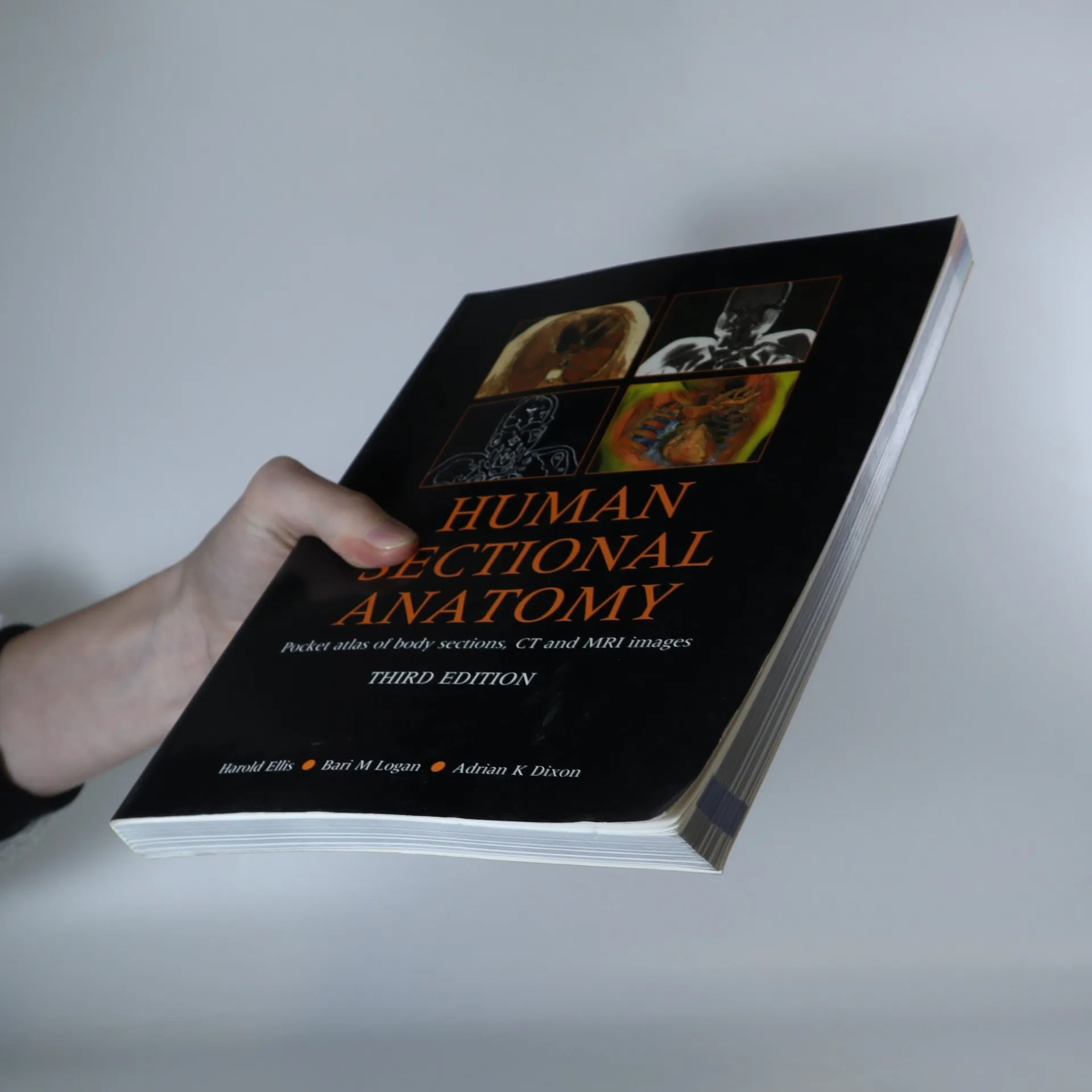
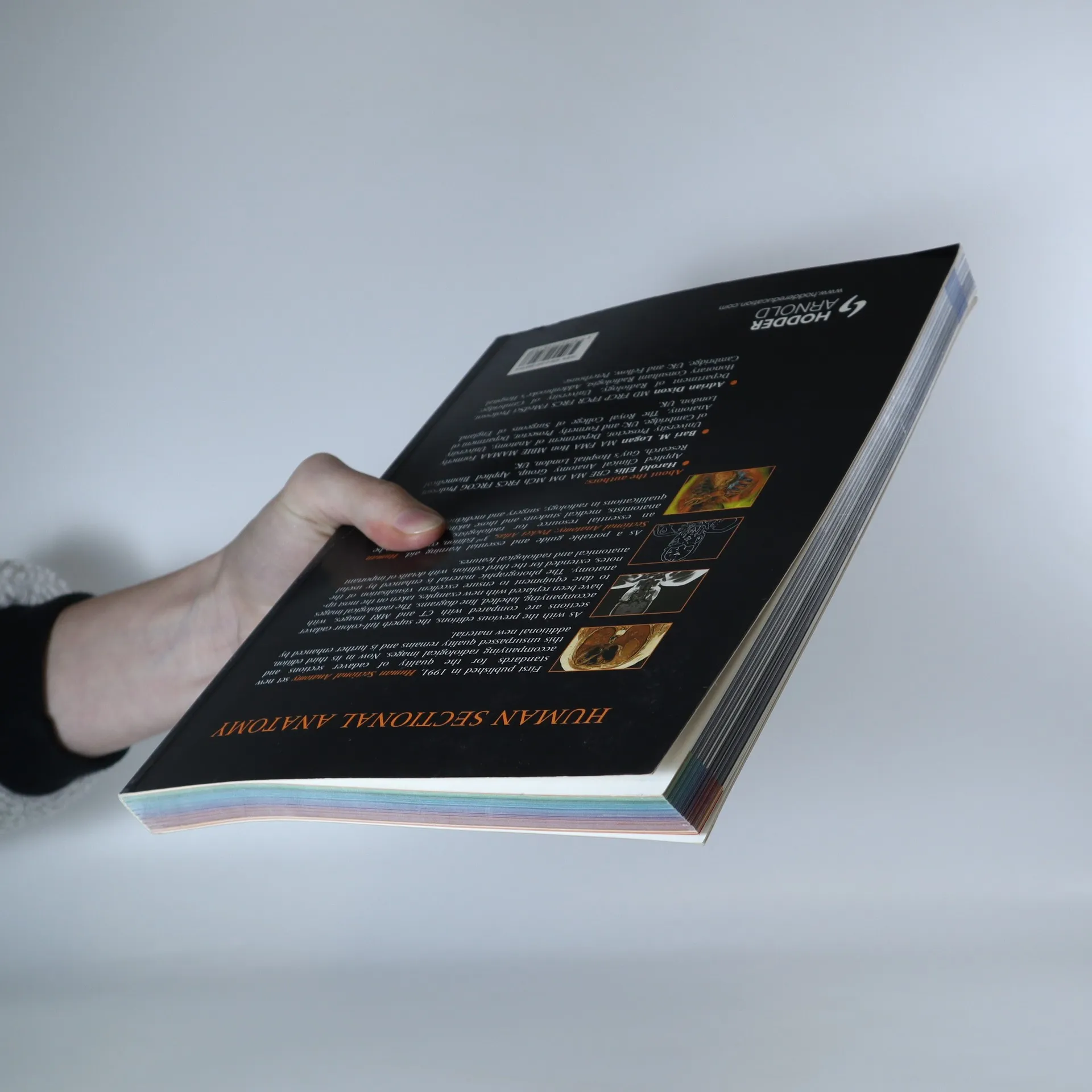
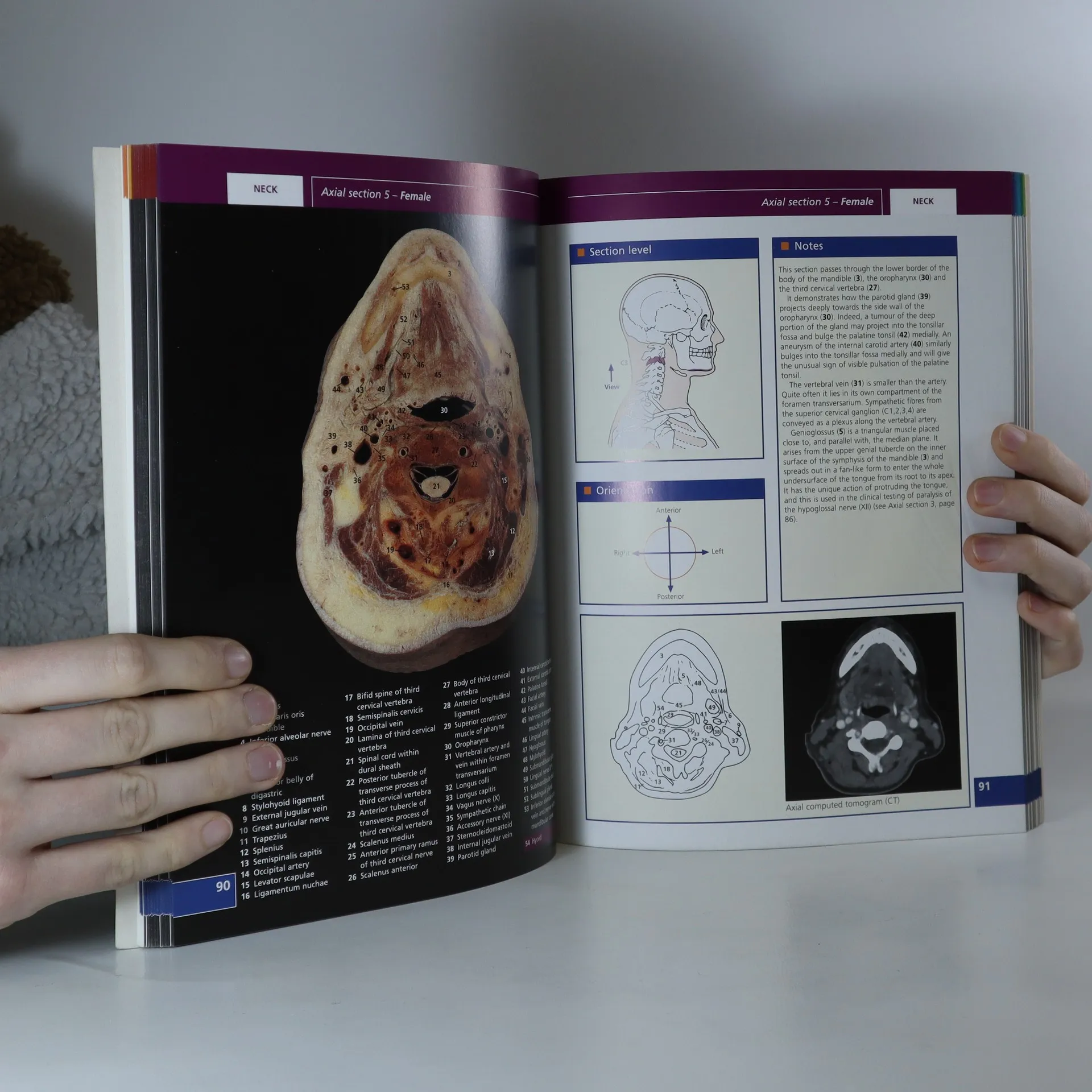
First published in 1991, Human Sectional Anatomy set new standards for the quality of cadaver sections and accompanying radiological images. Now in its third edition, this unsurpassed quality remains and is further enhanced by some useful new material. As with the previous editions, the superb full-colour cadaver sections are compared with CT and MRI images, with accompanying, labelled line diagrams. Many of the radiological images have been replaced with new examples, taken on the most up-to date equipment to ensure excellent visualisation of the anatomy. Completely new page spreads have been added to improve the book's coverage, including images taken using multidetector CT technology, and some beautiful 3D volume rendered CT images. The photographic material is enhanced by useful notes, extended for the third edition, with details of important anatomical and radiological features.
Zakup książki
Human sectional anatomy. Pocket atlas of body sections, CT and MRI images, Harold Ellis, Adrian K. Dixon, B. M. Logan
- Język

- Rok wydania
- 2009
- Oprawa
- (miękka)
Metody płatności
Brakuje nam tutaj Twojej recenzji.
- Tytuł
- Human sectional anatomy. Pocket atlas of body sections, CT and MRI images
- Język
- angielski
- Autorzy
- Harold Ellis, Adrian K. Dixon, B. M. Logan
- Wydawca
- Hodder Arnold
- Rok wydania
- 2009
- Oprawa
- miękka
- Liczba stron
- 267
- ISBN10
- 034098516x
- ISBN13
- 9780340985168
- Seria
- Tagi
- Atlasy
- Ocena
- 4,35 z 5
- Opis
- First published in 1991, Human Sectional Anatomy set new standards for the quality of cadaver sections and accompanying radiological images. Now in its third edition, this unsurpassed quality remains and is further enhanced by some useful new material. As with the previous editions, the superb full-colour cadaver sections are compared with CT and MRI images, with accompanying, labelled line diagrams. Many of the radiological images have been replaced with new examples, taken on the most up-to date equipment to ensure excellent visualisation of the anatomy. Completely new page spreads have been added to improve the book's coverage, including images taken using multidetector CT technology, and some beautiful 3D volume rendered CT images. The photographic material is enhanced by useful notes, extended for the third edition, with details of important anatomical and radiological features.