

Więcej o książce
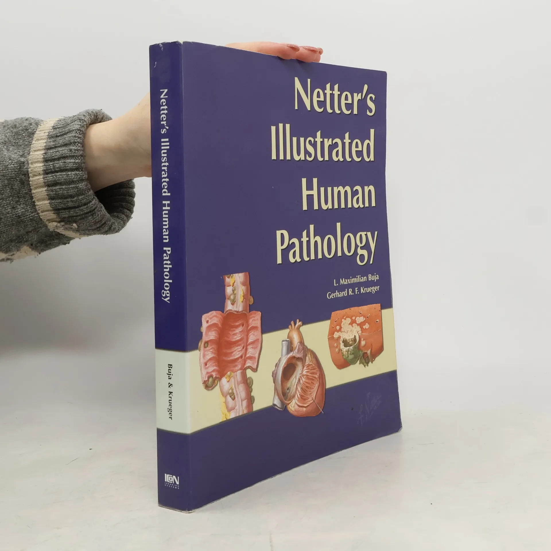
Netter's Atlas of Human Pathology is a visually vibrant approach to help students gain critical insight into the structure-function relationships and the pathological basis of human disease. It provides clear and succinct representations of common human diseases by relating anatomical changes to the functional and clinical manifestations of disease and their underlying causes and mechanisms. Features more than 380 classic Netter and new Netter-sytle images, gross and microscopic photographs and tables.The book consists of 12 organ-specific chapters - each containing precisely rendered illustrations of pathological processes and diseases accompanied by relevant text to expand knowledge and clarify understanding. Comparative data about similar disease processes are summarized in tables.Netter's Illustrated Human Pathology The Atlas offers a superb complement to more comprehensive textbooks and presentations of pathology, including course syllabi. It can also be used as an adjunct for study of gross and microscopic pathology specimens in laboratory exercises, and makes a great review resource for students, medical residents, physicians and other healthcare professionals.
Zakup książki
Netter's Illustrated Human Pathology, Gerhard R. F. Krueger, L. Maximilian Buja, Frank H. Netter
- Język

- Rok wydania
- 2004
- Oprawa
- (miękka)
Metody płatności
Brakuje nam tutaj Twojej recenzji.
- Tytuł
- Netter's Illustrated Human Pathology
- Język
- angielski
- Wydawca
- Saunders
- Rok wydania
- 2004
- Oprawa
- miękka
- Liczba stron
- 527
- ISBN10
- 1929007450
- ISBN13
- 9781929007455
- Seria
- Tagi
- Tematyka medyczna, Atlasy, Patologia
- Ocena
- 4,45 z 5
- Opis
- Netter's Atlas of Human Pathology is a visually vibrant approach to help students gain critical insight into the structure-function relationships and the pathological basis of human disease. It provides clear and succinct representations of common human diseases by relating anatomical changes to the functional and clinical manifestations of disease and their underlying causes and mechanisms. Features more than 380 classic Netter and new Netter-sytle images, gross and microscopic photographs and tables.The book consists of 12 organ-specific chapters - each containing precisely rendered illustrations of pathological processes and diseases accompanied by relevant text to expand knowledge and clarify understanding. Comparative data about similar disease processes are summarized in tables.Netter's Illustrated Human Pathology The Atlas offers a superb complement to more comprehensive textbooks and presentations of pathology, including course syllabi. It can also be used as an adjunct for study of gross and microscopic pathology specimens in laboratory exercises, and makes a great review resource for students, medical residents, physicians and other healthcare professionals.